Echocardiography: A Complete Guide to Heart Ultrasound
What is Echocardiography?
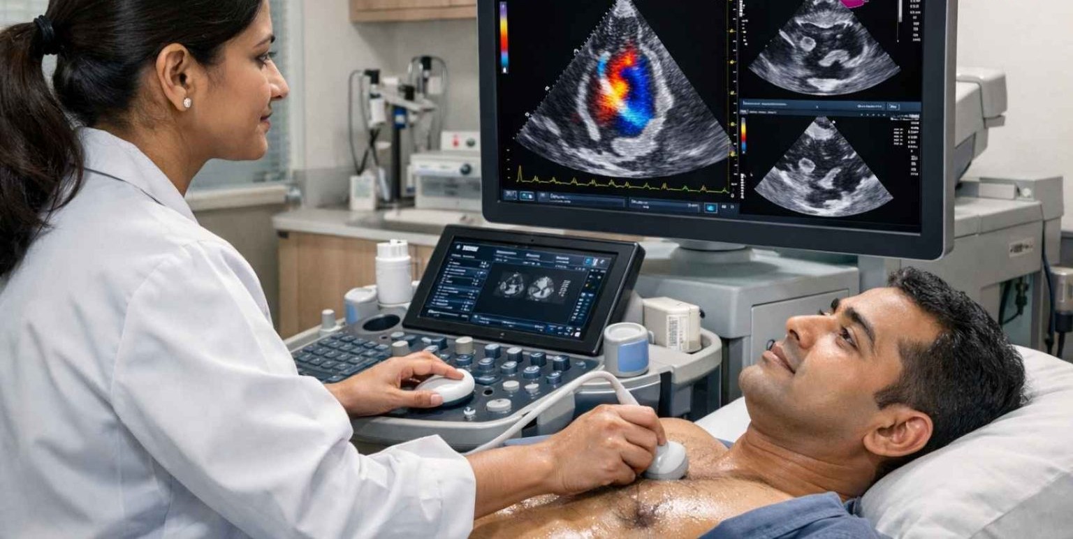
Echocardiography (Echo) is a non-invasive ultrasound test used to evaluate the structure and function of the heart. It uses sound waves to create real-time images of the heart, helping doctors diagnose and monitor various heart conditions.
It is one of the most commonly used and safest tests in cardiology.
Why is Echocardiography Done?
Echocardiography helps in:
- Assessing heart function
- Detecting heart valve diseases
- Diagnosing congenital heart defects
- Evaluating heart muscle health
- Identifying fluid around the heart (pericardial effusion)
- Monitoring heart conditions over time
Types of Echocardiography
1. Transthoracic Echocardiography (TTE)
- Most common type
- Probe placed on the chest
- Non-invasive and painless
2. Transesophageal Echocardiography (TEE)
- Probe inserted through the esophagus
- Provides more detailed images
- Used in complex cases
3. Stress Echocardiography
- Performed during or after exercise
- Assesses heart function under stress
4. Doppler Echocardiography
Measures blood flow and pressure within the heart
5. 3D Echocardiography
Provides detailed three-dimensional images
How is Echocardiography Performed?
- The patient lies on an examination table
- A gel is applied to the chest
- A transducer (probe) is moved over the chest
- Sound waves create images displayed on a monitor
- The procedure usually takes 15–30 minutes
Benefits of Echocardiography
- Non-invasive and painless
- No radiation exposure
- Provides real-time heart images
- Safe for all age groups
- Helps in early diagnosis of heart conditions
Risks and Safety
Echocardiography is extremely safe:
- No known side effects for standard echo
- TEE may cause mild throat discomfort
- Stress echo may involve slight exertion
What Conditions Can Echocardiography Detect?
- Heart valve diseases
- Heart failure
- Congenital heart defects
- Cardiomyopathy
- Blood clots in the heart
- Infections (endocarditis)
Preparation for Echocardiography
- No special preparation needed for standard echo
- Fasting may be required for TEE
- Comfortable clothing is recommended
When to Get an Echocardiogram?
Your doctor may recommend it if you have:
- Chest pain
- Shortness of breath
- Heart murmur
- Palpitations
- Swelling in legs
- History of heart disease
Our Specialist

Dr. Virender Singh Bhati
MD Physician, Clinical Cardiologist, PGDCC (SPS Apollo Hospital, Ludhiana)
Associate Consultant - Department of Cardiology, Fortis Hospital, Greater Noida